Get Bladder Carcinoma In Situ Images US
Get Bladder Carcinoma In Situ Images US. Carcinoma in situ may occur either close to or remote from an exophytic lesion or, rarely, it may occur as focal or diffuse lesions in a patient without macroscopic tumors. Bladder carcinoma often manifests at an early stage, and no imaging evaluation is performed to determine whether there are locoregional or systemic metastases.
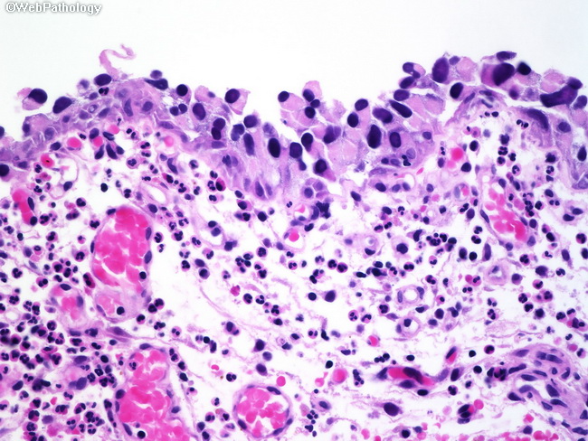
Stage 0is (also called carcinoma in situ) is a flat tumor on the tissue lining the inside of the bladder.
The subtypes of carcinomas include adenocarcinoma, squamous cell carcinoma, transitional cell carcinoma (in the bladder or kidneys), and basal cell. For other types of carcinoma in situ, routine screening and laboratory tests can detect precancer at an early stage. Brady urology at johns hopkins hospital. Tis or carcinoma in situ refers to nonpapillary (flat).
Posting Komentar untuk "Get Bladder Carcinoma In Situ Images US"